Should I Get a Cardiac MRI?
Medical Disclaimer
Always consult a licensed healthcare professional when deciding on medical care. The information presented on this website is for educational purposes only and exclusively intended to help consumers understand the different options offered by healthcare providers to prevent, diagnose, and treat health conditions. It is not a substitute for professional medical advice when making healthcare decisions.
Overview
Cardiac MRI represents a sophisticated, radiation-free imaging technology that has emerged as a comprehensive diagnostic tool for evaluating heart disease. Unlike traditional cardiac imaging methods, this advanced technique can assess multiple aspects of heart health in a single examination, including function, blood flow, tissue viability, and structural abnormalities. The technology has proven highly effective across diverse clinical applications, with studies demonstrating exceptional accuracy in detecting coronary artery disease, predicting surgical outcomes, and diagnosing complex heart conditions.
The greatest beneficiaries include patients with unexplained chest pain despite normal coronary angiograms, those considering heart procedures who need viability assessment, individuals with heart failure or suspected cardiomyopathy, and patients with inconclusive results from other imaging tests. Doctors typically prescribe cardiac MRI when comprehensive cardiac evaluation is needed, particularly for complex diagnostic challenges or treatment planning decisions.
Cardiac MRI maintains an excellent safety profile with minimal risks, primarily related to device compatibility and rare contrast reactions rather than the procedure itself. The scans are performed in specialized imaging facilities equipped with appropriate MRI technology and trained cardiac imaging specialists.
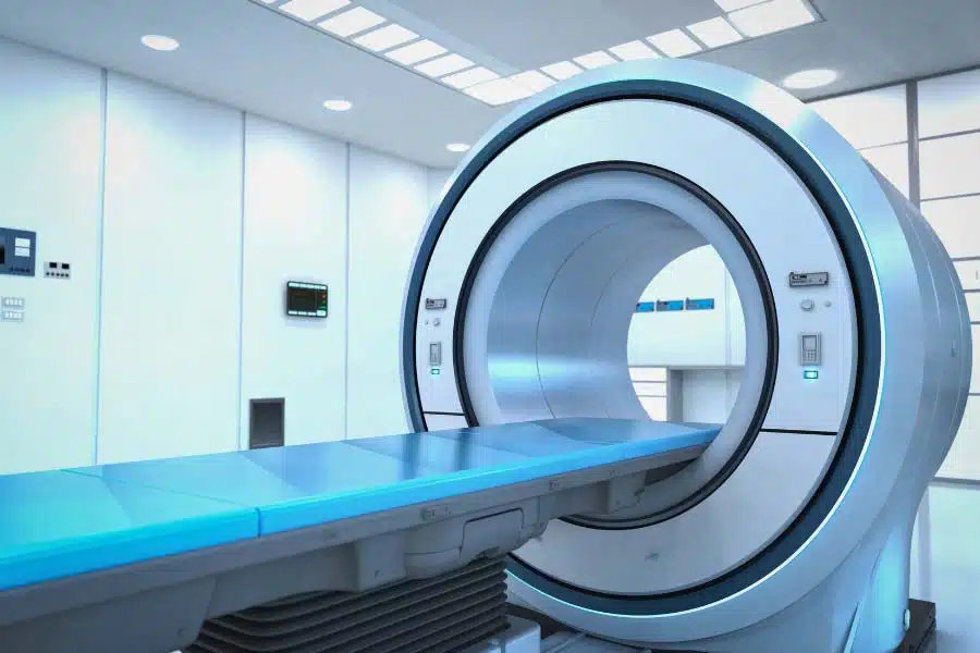
What is a cardiac MRI?
Cardiac MRI (also called cardiac magnetic resonance imaging or CMR) is a sophisticated, non-invasive imaging technique that uses powerful magnetic fields and radio waves to create detailed pictures of the heart and surrounding blood vessels. Unlike other cardiac imaging methods, cardiac MRI provides superior spatial resolution and can comprehensively assess multiple aspects of cardiac health in a single examination without using ionizing radiation (Poon et al., 2002).
The technology excels at evaluating cardiac function, myocardial perfusion, tissue viability, and even coronary anatomy all in one session. Cardiac MRI can detect scar tissue through late gadolinium enhancement imaging, which helps identify areas of previous heart damage and predict which patients might benefit from procedures like coronary revascularization (Kim et al., 2000). The technique also enables quantitative stress perfusion imaging that can accurately diagnose coronary artery disease and distinguish between blockages in large coronary arteries versus problems with the heart’s smallest blood vessels (Catania et al., 2025).
What makes cardiac MRI particularly valuable is its ability to serve as a “one-stop-shop” evaluation for various heart conditions, offering detailed functional and structural information that often eliminates the need for multiple separate tests or more invasive procedures (Poon et al., 2002). The technology continues to advance with innovations like deep learning-based image reconstruction that can significantly reduce scan times while maintaining diagnostic quality (Klemenz et al., 2024).
Does a cardiac MRI work?
Yes, cardiac MRI has proven highly effective across numerous clinical applications. Studies demonstrate that quantitative cardiac MRI perfusion measures achieve significantly higher accuracy with area under the curve values of 0.90 and 0.88 in detecting coronary microvascular dysfunction compared to visual assessment alone (Rahman et al., 2021). For diagnosing coronary artery disease more broadly, combined cardiac MRI protocols reach 87% accuracy in detecting significant coronary stenosis when compared against invasive coronary angiography (de Mello et al., 2012).
The technology excels particularly in myocardial viability assessment, where contrast-enhanced MRI can predict improvement in regional ventricular contraction after coronary revascularization based on the transmural extent of scar tissue (Kim et al., 2000). This predictive capability has proven so reliable that cardiac MRI should be considered a first-line test in assessing myocardial viability when making revascularization decisions (Al-Sabeq et al., 2019).
Beyond coronary disease, cardiac MRI demonstrates diagnostic value across various conditions. In cardiomyopathy evaluation, studies show cardiac MRI identified cardiomyopathy in 75% and arrhythmia in 25% of patients, demonstrating high positive predictive value for diagnosing various heart muscle diseases (Singh et al., 2024). The technology’s effectiveness extends even to complex diagnostic challenges like distinguishing types of cardiac amyloidosis, where deep learning analysis of cardiac MRI outperformed experienced human readers in classification accuracy (Germain et al., 2023).
Discover the tests and treatments that could save your life
Get our unbiased and comprehensive report on the latest techniques for heart disease prevention, diagnosis, and treatment.
Why get a cardiac MRI?
Cardiac MRI offers several compelling advantages that make it valuable for both diagnosis and treatment planning. The primary reason is its ability to provide comprehensive cardiac assessment without radiation exposure, combining multiple evaluations that would otherwise require separate tests.
For patients with chest pain and normal-looking coronary arteries, cardiac MRI can identify coronary microvascular dysfunction that other tests might miss, helping explain symptoms when traditional coronary angiography appears normal (Rahman et al., 2021). This is particularly important since microvascular disease can cause significant symptoms despite the absence of major coronary blockages.
When considering heart procedures like bypass surgery or stenting, cardiac MRI provides crucial information about myocardial viability to predict which patients will benefit from revascularization. The scan can show which areas of heart muscle are still alive versus scarred, helping doctors and patients make informed decisions about whether invasive procedures are likely to improve heart function (Kim et al., 2000).
For patients with unexplained heart failure or irregular heart rhythms, cardiac MRI serves as a powerful diagnostic tool that can enhance clinical decision-making by identifying the underlying cause of cardiomyopathy (Singh et al., 2024). The detailed tissue characterization capabilities allow doctors to distinguish between different types of heart muscle disease, which can dramatically impact treatment approaches.
The technology’s comprehensive nature means that cardiac MRI can serve as a central tool to evaluate ischemic heart disease more efficiently than traditional imaging approaches, potentially avoiding the need for multiple separate tests (Poon et al., 2002).
Who most benefits from a cardiac MRI?
Several specific patient groups derive the greatest benefit from cardiac MRI based on the technology’s unique diagnostic capabilities.
Patients with chest pain but normal-appearing coronary arteries represent a key beneficiary group, as cardiac MRI can detect coronary microvascular dysfunction that explains their symptoms when other tests appear normal (Rahman et al., 2021). This is particularly valuable since quantitative stress perfusion cardiac MRI improves ischemia detection and management, especially in patients with angina and nonobstructive coronary artery disease (Catania et al., 2025).
Individuals with heart failure or suspected cardiomyopathy benefit significantly, as cardiac MRI should be considered a first-line diagnostic tool for evaluating patients with arrhythmia or unexplained cardiomyopathy (Singh et al., 2024). The technology excels at tissue characterization, even enabling distinction between different types of cardiac amyloidosis that can dramatically impact treatment approaches (Germain et al., 2023).
Patients considering revascularization procedures like bypass surgery or stenting represent another crucial group. Cardiac MRI can guide revascularization decisions in patients with impaired ventricular function by identifying viable myocardium, helping determine who will actually benefit from these invasive procedures (Child and Das, 2012). The likelihood of functional improvement after revascularization declines as the transmural extent of scar tissue increases, making this assessment critical for treatment planning (Kim et al., 2000).
Those with inconclusive results from other imaging tests also benefit, as cardiac MRI often provides additional clarity when other modalities fall short in diagnosing ischemic heart disease (Sawlani and Collins, 2016).
How does a cardiac MRI compare to other imaging methods when diagnosing or treating coronary artery disease?
Cardiac MRI offers several distinct advantages over other imaging modalities in coronary artery disease evaluation, particularly in its comprehensive assessment capabilities and diagnostic accuracy. Studies demonstrate that combined cardiac MRI protocols reach 87% accuracy in detecting significant coronary stenosis when compared against invasive coronary angiography, which serves as the gold standard (de Mello et al., 2012).
One of cardiac MRI’s key strengths lies in its ability to detect problems that other imaging methods might miss. For patients with chest pain but normal-appearing coronary arteries on angiography, cardiac MRI can identify coronary microvascular dysfunction that explains persistent symptoms when traditional tests appear normal (Rahman et al., 2021).
In viability assessment, cardiac MRI demonstrates superior predictive capabilities compared to other modalities. The technology can predict improvement in regional ventricular contraction after coronary revascularization based on the transmural extent of scar tissue, providing more precise guidance for treatment decisions than many alternative imaging approaches (Kim et al., 2000). This has led experts to conclude that cardiac MRI should be considered a first-line test in assessing myocardial viability when making revascularization decisions (Al-Sabeq et al., 2019).
Unlike nuclear imaging or CT angiography, cardiac MRI provides this comprehensive evaluation without ionizing radiation exposure. The technology can serve as a central tool to evaluate ischemic heart disease more efficiently than traditional imaging approaches, potentially eliminating the need for multiple separate tests (Poon et al., 2002).
When do doctors typically prescribe a cardiac MRI?
Doctors typically order cardiac MRI in several specific clinical scenarios where its unique capabilities provide critical diagnostic information.
When patients present with chest pain but coronary angiography shows no significant blockages, doctors often turn to cardiac MRI to identify coronary microvascular dysfunction that could explain persistent symptoms (Rahman et al., 2021). This is particularly common when traditional stress tests or other imaging methods haven’t provided clear answers.
Before major heart procedures, cardiac MRI is frequently ordered to assess tissue viability. Doctors use it to determine which patients with impaired ventricular function might benefit from revascularization by identifying areas of viable versus scarred heart muscle (Child and Das, 2012). This assessment is crucial since the likelihood of functional improvement after revascularization depends heavily on the extent of scar tissue present (Kim et al., 2000).
When patients have unexplained heart failure, irregular heart rhythms, or suspected heart muscle disease, doctors often prescribe cardiac MRI as a first-line diagnostic tool for evaluating cardiomyopathy (Singh et al., 2024). The detailed tissue characterization helps distinguish between different causes of heart muscle problems, which can dramatically impact treatment decisions.
Cardiac MRI is also commonly ordered when other imaging tests are inconclusive or insufficient for making a definitive diagnosis in ischemic heart disease (Sawlani and Collins, 2016). Its comprehensive assessment capabilities often provide the additional clarity needed when echocardiography or nuclear stress tests haven’t yielded clear results.
What are the risks of a cardiac MRI?
Cardiac MRI is generally considered a very safe procedure with minimal risks, particularly when compared to invasive alternatives or radiation-based imaging. The uploaded sources emphasize safety as one of cardiac MRI’s key advantages, with one review noting that cardiac MRI should be considered a first-line test in assessing myocardial viability, given its safety compared to other diagnostic approaches (Al-Sabeq et al., 2019).
The primary risks are related to contraindications rather than direct procedural complications. Patients with certain implanted devices may not be candidates for cardiac MRI unless their devices are specifically MRI-compatible. Some patients may experience claustrophobia due to the enclosed nature of the MRI scanner, though this can often be managed with sedation or open MRI systems where available.
When contrast agents are used, there are rare risks of allergic reactions to gadolinium-based contrast materials. Additionally, patients with severe kidney dysfunction face a small risk of nephrogenic systemic fibrosis, a rare condition associated with gadolinium contrast, though this has become increasingly uncommon with newer contrast agents and careful patient screening.
The safety profile of cardiac MRI has contributed to its growing adoption as a preferred diagnostic tool. The absence of ionizing radiation makes it particularly attractive for patients who might otherwise require multiple imaging studies or for those where radiation exposure is a concern. Recent technological advances, including low-field cardiac MRI systems, may further improve accessibility and reduce contraindications for patients with certain implants (Campbell-Washburn et al., 2024).
Don’t become a statistic
Learn the latest strategies to prevent, diagnose, and manage heart disease in our one-of-a-kind report.
Where are cardiac MRIs performed?
Cardiac MRIs are performed in specialized medical imaging facilities that have the necessary equipment and expertise to conduct these sophisticated scans. The procedure requires powerful MRI machines specifically configured for cardiac imaging, along with trained technologists and radiologists who specialize in cardiac MRI interpretation.
Most cardiac MRIs are conducted in hospital-based imaging departments, dedicated cardiac imaging centers, or specialized radiology facilities. These locations must have the appropriate MRI hardware, which traditionally includes high-field strength magnets (typically 1.5 Tesla or 3 Tesla systems). However, recent advances suggest that low-field cardiac MRI could increase accessibility and potentially be performed in more diverse settings due to reduced infrastructure requirements and costs (Campbell-Washburn et al., 2024).
The facilities performing cardiac MRI must also have specialized software and protocols for cardiac imaging sequences, as well as the capability to administer contrast agents safely when needed. Additionally, they require staff trained specifically in cardiac MRI techniques, as the imaging protocols and interpretation differ significantly from routine body MRI scans.
Some research centers and academic medical institutions are exploring even more advanced applications, with studies demonstrating the feasibility of routine cardiac MRI at 7 Tesla using commercially available hardware, though such ultra-high-field systems remain primarily in research settings (Reiter et al., 2021).
The location where you receive your cardiac MRI will depend on your healthcare provider’s referral patterns, insurance coverage, and the availability of cardiac MRI services in your geographic area.
What is the cost of a cardiac MRI, and who pays for it?
The cost of cardiac MRI typically ranges from $1,000 to $5,000, with significant variation based on geographic location, facility type, and specific imaging protocols required. The exact cost depends on factors such as whether contrast agents are used, the complexity of the study, and regional healthcare pricing differences.
Insurance coverage for cardiac MRI is generally good when the procedure is deemed medically necessary by your physician. Most major insurance plans, including Medicare, typically cover cardiac MRI for appropriate clinical indications such as evaluating chest pain, assessing heart function, or determining treatment options for heart disease. However, coverage policies can vary between insurers, so it’s important to verify your specific benefits before scheduling the procedure.
Patients may still be responsible for deductibles, copayments, or coinsurance amounts even when the procedure is covered. For those without insurance coverage or with high-deductible plans, the out-of-pocket costs can be substantial. Some facilities offer payment plans or financial assistance programs to help manage these expenses.
The potential for low-field cardiac MRI systems to reduce costs while maintaining diagnostic quality could make cardiac MRI more accessible in the future (Campbell-Washburn et al., 2024). These technological advances might help address cost barriers that currently limit access to cardiac MRI for some patients.
Before scheduling your cardiac MRI, it’s advisable to contact both your healthcare provider’s office and your insurance company to understand your expected out-of-pocket costs and ensure proper prior authorization if required.
Conclusion
Cardiac MRI represents one of several advanced imaging options available within the modern cardiac care ecosystem. This technology provides a non-invasive approach to comprehensive heart evaluation that can offer valuable diagnostic information across a range of cardiac conditions. The technique’s ability to assess multiple cardiac parameters in a single examination, combined with its strong safety profile, positions it as a significant tool in contemporary cardiology practice.
Understanding the capabilities, applications, and limitations of cardiac MRI helps inform the broader conversation between patients and healthcare providers about appropriate diagnostic approaches. As cardiac imaging technology continues to evolve, cardiac MRI stands as an example of how advanced medical imaging can contribute to more precise diagnosis and treatment planning in cardiovascular medicine.